
Is the whole is greater than the sum of the parts?
Required Reading - Chapter 8, Simmons & Young
How are neurons assembled
to form networks?
 |
 |
|
|
|
Aplysia II (the sequel)
Aplysia is a sea-slug like Tritonia. It breathes
by drawing water over a gill, which it extends from its body.
The gill is retracted in response to touch, and is also retracted
in time with respiratory pumping movements.
Control of gill movements
Some sensory neurons with receptors in the skin close to the gill
make direct, exitatory synapses onto gill withdrawal motor neurons.
An increase in stimulus strength leads to increase in sensory
neuron firing rate, which leads in turn to increase in the intensity
of the motor neuron response. The spatial and temporal summation
of post-synaptic potentials in the motor neurons could
explain the relationship between stimulus intensity and response.
But...
Estimates of the contribution of these direct circuits to the
gill withdrawal response range from 5% to 60%. Sensory neurons
also make synapses with interneurons, which in turn synapse with
motor neurons. This forms a parallel arrangement of direct
and indirect pathways for information to flow from sensory system
to motor system - similar to the flight motor pattern generator
in locusts.
Optical monitoring of neuronal
activity
Ganglia are injected with a dye that changes spectral properties
depending on the voltage of the cell membrane. Light is shone
through the ganglion, and the amount of light that passes through
is recorded. Changing neuronal activity shows up as changing patterns
of light transmission. Although this technique is very useful,
it does not allow recording from individual identified neurons.
New interpretation of gill movements
Three kinds of movements were studied:
It turned out that hundreds of neurons in the abdominal ganglion
are active during gill movements. 62 - 72 neurons were usually
active during each movement, and almost all of these were active
during all three kinds of movement.
This seems to be indicative of distributed circuits
Leg reflex movements in the locust

When you touch a locust on the leg it moves the leg away from the point of contact. These movements usually involve several joints. The touch is detected by several types of sensilla:
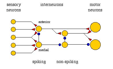
Sensory neurons project to form a somatotopic map within
the nervous system, which means that specific locations on the
body are represented by particular parts of the nervous system.
The reflex is controlled by neurons in the 3rd thoracic ganglion.
Levels of information processing in leg reflex
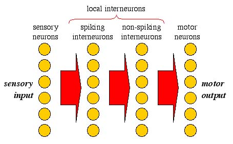
local spiking interneurons
Each spiking interneuron has two layers of branches:
There are two main groups of spiking interneurons:

Each spiking interneuron is responds to sensilla within a specific region of the leg and shows rapid adaptation to sensory input. Each spiking interneuron synapses with a umber of targets, including non-spiking interneurons and motor neurons. They form a diffuse pattern of connectivity, but somatotopic information is retained.
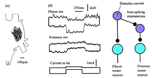
local non-spiking interneurons
These interneurons provide the main input to motor neurons. These
neurons do not generate action potentials - instead, changes
in membrane potential directly affect the rate of neurotransmitter
release. There is a linear relationship between membrane potential
in interneuron and motor neuron. Non-spiking interneurons receive
input from many sensory cells via spiking interneurons. They act
as summation points for information about posture - provide behavioural
context for movements. For example, they can be more sensitive
to increase or decrease in joint angle, providing information
about the direction of movement.
Organisation of neurons in leg reflexes
Some interesting comparisons and take-home messages
The last few examples have been of diffuse, distributed circuits
of neurons.
How does this compare with the dedicated circuits seen in the
escape mechanisms covered earlier in the course?
The reflexes in locust legs take information from spiking
sensory neurons and convert it into graded membrane potentials.
How does this compare with the way information is processed
in the visual system?